Research
2-dimensional pattern classification
This research was done for my Master's thesis, under the supervision of Dr. Patrick Shipman.
Biological systems are flush with organized patterns, caused by a variety of underlying mechanisms on multiple different scales. Alan Turing, primarily known for his work in computer science, was also one of the first mathematicians to propose a mathematical model for biological pattern formation in The Chemical Basis of Morphogenesis.
Patterns commonly occur in the petals of flowers, where spatial variation in pigment concentration causes a variety of spectacular patterns. Flower petal patterns are known to play a role in pollinator foraging activity. One genus of flowers known for distinctive patterns is the monkeyflowers, Mimulus. Previous work by Ding and colleagues has shown that the M. lewisii anthocyanin synthesis system satisfies a model for spontaneous pattern formation proposed by Gierer and Meinhardt. As a result, it's possible to simulate images that resemble Mimulus patterns.
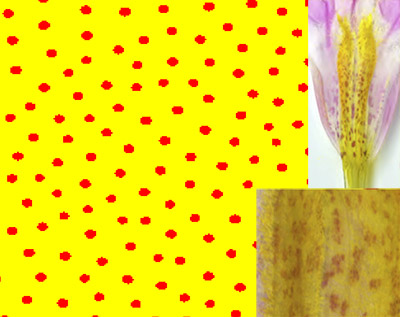
Pattern classification using machine learning is a topic of interest because patterns can tell us a lot about the conditions that formed them. However, classifying patterns from photos is a challenge because many models operate in 3 dimensions, while patterns only offer 2-dimensional information. Since simulated data from the model is needed to train the classifier, something needs to be done to bridge the gap between the 3-D and 2-D data. My thesis focused on developing an algorithm that can reliably convert a binary 2-dimensional slice from the 3-D data into a set of points, then use topological data analysis and machine learning on those points to train a classifier. I also explored the effectiveness of the machine learning model on binarized versions of real flower photos.
Machine learning for EIT
This research was done under the supervision of Dr. Jennifer Mueller and in a group with Kyler Howard, Tyler Stephens, and Natalie Wijesinghe.
Electrical Impedance Tomography (EIT) is a noninvasive medical imaging technique using electric fields. Like CT, it can be used to get images of the lungs. EIT has many advantages over CT, namely that it can be used frequently and in real time and doesn't require the use of radiation. However, the image quality is poor compared to CT. In this project, we segmented CT scans and used the segmentations to generate simulated EIT voltages (the quantities being measured) and conductivities (the quantities that are used in image reconstruction) for training data for a neural network. This neural network takes voltage measurements as input directly, then outputs conductivities that can be viewed as an image, so our goal is to use it along with the EIT hardware to improve images in real time.
This work has been published in the Journal of Computational and Applied Mathematics!